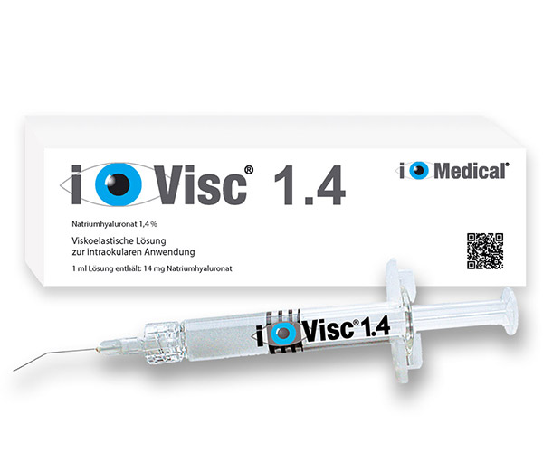
Ophthalmic Viscosurgical Devices (OVDs) are gel-like substances used in various ophthalmic surgeries to maintain space, protect tissues, and facilitate surgical maneuvers. Their primary applications include:
1. Cataract Surgery (Phacoemulsification)
OVDs play a crucial role in modern cataract surgery by:
Creating and maintaining anterior chamber space → Prevents corneal collapse during incisions.
Protecting corneal endothelial cells → Shields from ultrasound energy and mechanical trauma.
Facilitating intraocular lens (IOL) implantation → Helps position the IOL accurately in the capsular bag.
Stabilizing the capsular bag → Prevents posterior capsule rupture.
Common OVDs Used:
Cohesive OVDs (e.g., i.Visc Healon®, Healon GV®) – Ideal for maintaining space.
Dispersive OVDs (e.g., i.Visc, Viscoat®, Ocucoat®) – Better for endothelial protection.
Viscoadaptive OVDs (e.g., Healon5®) – Adjust viscosity based on shear force.
2. Glaucoma Surgery (Trabeculectomy & Tube Shunts)
OVDs assist in:
Maintaining bleb formation → Helps in filtration surgeries.
Preventing intraoperative hypotony → Stabilizes anterior chamber depth.
Reducing scarring → Some OVDs may have anti-fibrotic effects.
3. Corneal Transplant (Penetrating & Endothelial Keratoplasty)
Deep Anterior Lamellar Keratoplasty (DALK) → OVDs help separate corneal layers.
Descemet’s Stripping Automated Endothelial Keratoplasty (DSAEK) → Protects donor tissue during insertion.
Descemet Membrane Endothelial Keratoplasty (DMEK) → Aids in unscrolling the graft.
4. Retinal Surgery (Vitrectomy & Detachment Repair)
Maintaining vitreous cavity space → Prevents collapse during fluid-air exchange.
Protecting the retina → Minimizes iatrogenic damage.
Assisting in silicone oil removal → Helps displace residual oil.
5. Pediatric & Complicated Cataract Surgery
Preventing anterior chamber collapse in small eyes (e.g., congenital cataracts).
Managing weak zonules → OVDs stabilize the capsular bag.
6. Secondary IOL Implantation & Refractive Lens Exchange
Sulcus-supported IOL placement → OVDs prevent iris prolapse.
Correcting aphakia → Helps position anterior or posterior chamber IOLs.
7. Other Uses
Intraocular pressure (IOP) management → Some OVDs help control postoperative IOP spikes.
Diagnostic procedures → Used in gonioscopy and anterior segment OCT imaging.
Trauma cases → Temporarily stabilizes the eye during repair.
Key Considerations in OVD Selection
Property Cohesive OVDs Dispersive OVDs Viscoadaptive OVDs Space Maintenance Excellent Poor (flows out) Excellent (adjustable) Endothelial Protection Moderate Excellent Excellent Ease of Removal Easy (single mass) Difficult (fragments) Moderate Best Use Case IOL implantation, capsular stability Phacoemulsification (endothelial protection) Complex cases (e.g., weak zonules)

